Scarred heart and clotting blood are winning scientific images in national competition

Judges’ winner - Stranger Strings: Platelets on the Hook
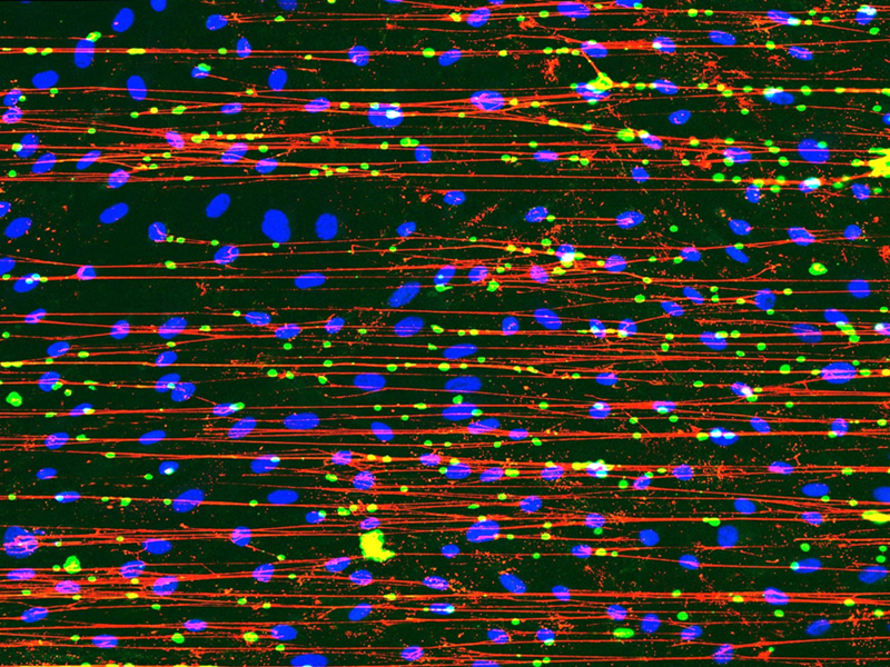
‘Stranger Strings’, a scientific snapshot of the first phase of blood clot formation, was submitted by BHF-funded researcher, Harriet Todd. The blue dots are cells which line the inside of blood vessels and release a sticky protein called von Willebrand factor (vWF).
This protein forms a net, pictured in red, to catch cells, known as platelets, which circulate in the blood. The platelets (shown in green) cause the blood to clot, which prevents excessive bleeding when someone is injured.
But researchers are looking at how this process can go wrong in people with diabetes, whose altered blood sugar control can trigger an excessive release of vWF. If left uncontrolled, this flood of vWF can increase the risk of a heart attack and stroke.
"Our project aims to string together the story of how microscopic processes in the body can leave us vulnerable to heart attacks and strokes. This image is a clear example of how science can be both visually stunning and essential to hopefully developing new treatments and saving lives.”
Supporters’ winner - The Heart's Silent Fight
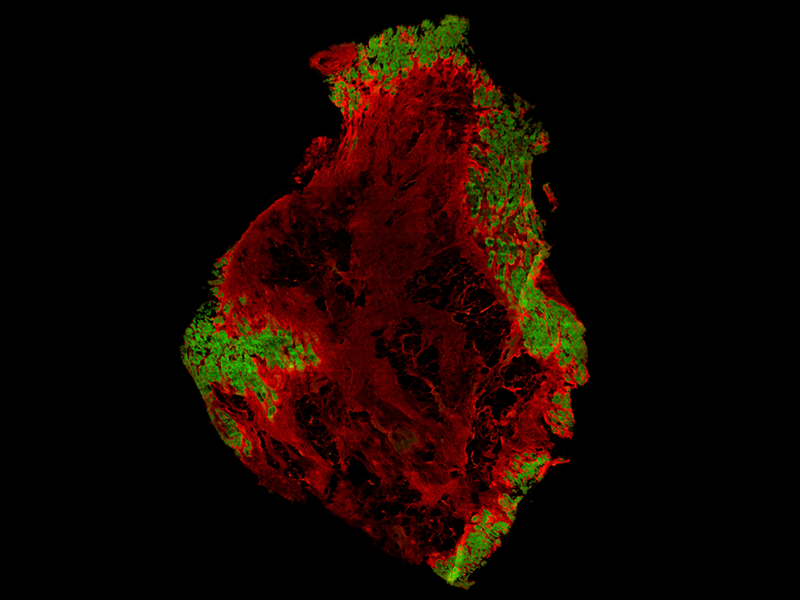
The Heart’s Silent Fight' captured the hearts and minds of our supporters.
The image, submitted by BHF-funded researcher Natalia Georgiadou, shows a human heart in the grip of fibrosis - the formation of scar tissue in the heart. Scarring develops when the heart muscle is damaged, often after a heart attack.
The scar tissue, shown in red within this image, cannot function like the normal heart cells, pictured in green. As a result, the heart’s ability to pump can be permanently impaired, meaning that some people develop heart failure. Researchers used coloured dyes to stain heart tissue, making healthy and scarred areas visible in the image. They aim to understand how healthy cells are replaced by scar tissue over time, and to find ways to stop this process, in order to help keep the heart beating normally.
Natalia Georgiadou said: “I am thrilled that this powerful image has resonated with supporters in the British Heart Foundation’s Reflections of Research competition. “We wanted to deliver a stark reminder of the damage a heart attack can leave behind. We hope our research will uncover the processes that turn healthy heart cells into scar tissue, to eventually try to stop heart failure before it strikes.”
'Bringing hope alive'
Dr Charmaine Griffiths, our Chief Executive, who was a competition judge, said: “Our Reflections of Research competition has now been running for 20 years, and I never fail to be amazed by the inventive ways in which our scientists showcase the beauty within the British Heart Foundation's lifesaving research.“The judges’ winning image so perfectly captures the remarkable ability of our bodies to heal themselves. The stunning art is not just beautiful, it also brings to life the hope we have in BHF research to make breakthroughs that save and improve countless lives.”
Dr Fraser Macrae, a BHF-funded Academic Fellow at the University of Leeds, a previous two-time winner of Reflections of Research and this year’s guest judge, said:
“This year’s entries offer a fascinating glimpse into the complex research led by BHF-funded scientists to understand more about the inner workings of our heart and circulatory systems in health and disease.
“I was amazed by the winning image, which shows how cells lining our blood vessels can release a sticky net to capture platelets that help to form a blood clot. This process is vital for repairing injuries to our blood vessels, and seeing it visualised so vividly makes you reflect on the hidden beauty and complexity of science.”
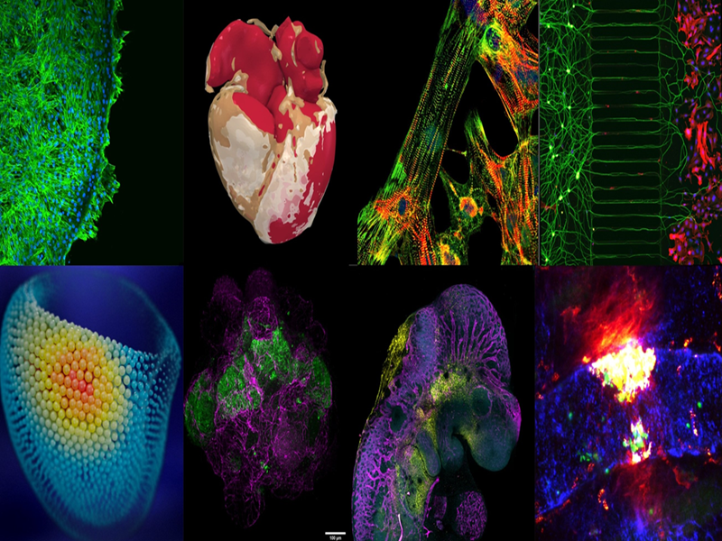
The remaining images on the shortlist are included in the below montage.