Reflections of Research
Find out more about our Reflections of Research imagery competition and how BHF-funded researchers can enter.
Our Reflections of Research competition began in 2005 and has been going strong ever since. Over the years, winning images have been features in national press, on the BBC and in exhibitions around the country, including in front of the London Eye.
Harriet Todd, based at the University of Leeds, submitted the captivating image below, winning over the judging panel to be named the 2025 Judges' Winner.
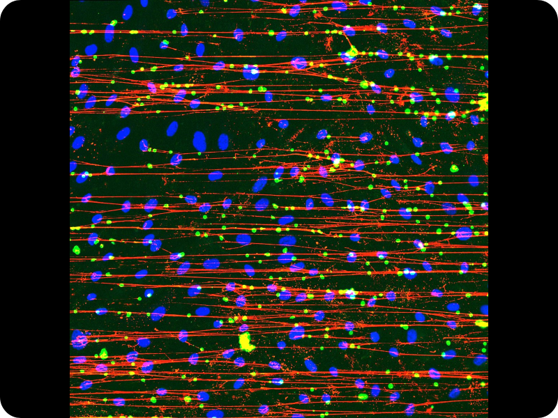
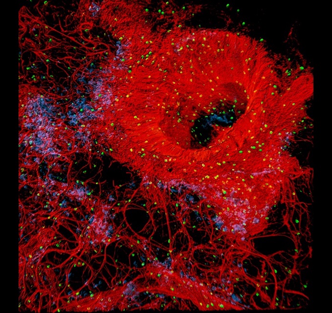
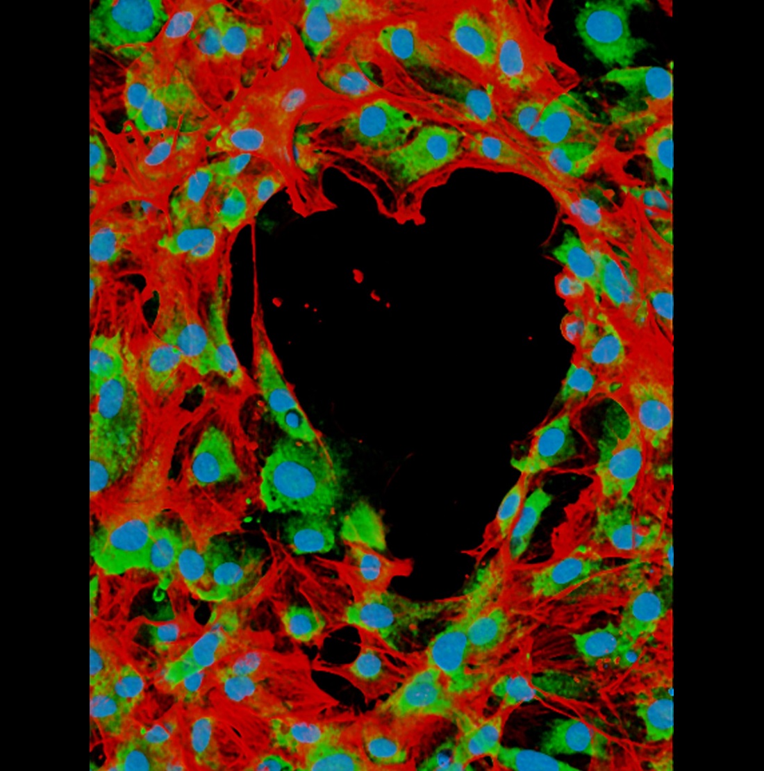
Harriet's image shows the first phase of blood clot formation. The blue circles are endothelial cells, from the inner lining of blood vessels. They have been provoked into releasing a protein, von Willebrand factor (VWF), which form the red strings. These strings attach to each other, creating a net that catches platelets (the green dots) from the circulation. This is the body’s natural response to injury to prevent excessive bleeding.
Harriet’s research investigates how this process can go wrong: endothelial cells are constantly in contact with blood and must be able to react appropriately to changes in blood sugar. This intricate response may be disrupted in people with diabetes. If the release of VWF is not controlled, it could contribute to injury and increase the risk of heart attack and stroke. The researcher aims to understand this process better to pave the way for new therapies.
Harriet said: “I am incredibly honoured to be the Judges’ Winner for the Reflections of Research competition this year. Our project aims to string together the story of how microscopic processes in the body can leave us vulnerable to heart attacks and strokes. This image is a clear example of how science can be both visually stunning and essential to hopefully developing new treatments and saving lives.”
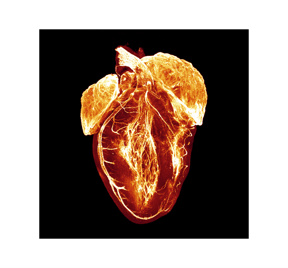
‘The Heart’s Silent Fight' captured hearts and minds, taking home the Supporters’ Favourite award.
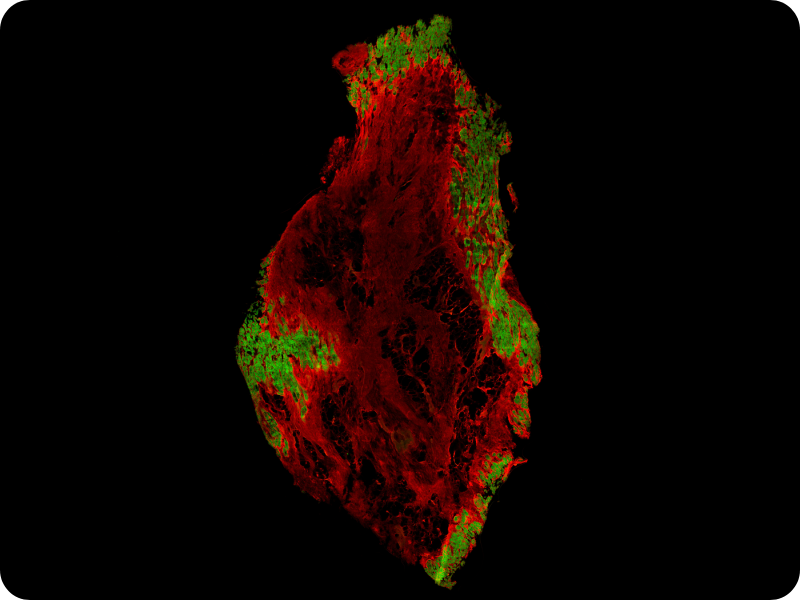
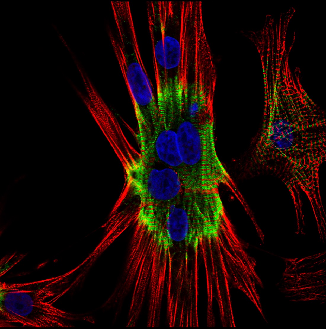
Natalia's image shows a human heart affected by fibrosis, where healthy heart tissue, shown in green, is replaced by scar tissue, shown in red. Scar tissue forms when the heart doesn’t get enough blood carrying oxygen, often after a heart attack, causing permanent damage. Scar tissue cannot function like normal heart cells, meaning the heart’s ability to pump blood is reduced, which can lead to heart failure in some cases.
To capture this image, Natalia used coloured dyes to stain the heart tissue, making healthy and scarred areas visible. The aim of her research is to understand how healthy heart cells are arranged in hearts impacted by fibrosis and how this changes as heart disease progresses. By doing so, she hopes to learn how healthy cells become replaced by scar tissue and find ways to stop this process in order to help keep the heart beating normally.
Natalia said: “I am thrilled that this powerful image has resonated with BHF supporters. We wanted to deliver a stark reminder of the damage a heart attack can leave behind. We hope our research will uncover the processes that turn healthy heart cells into scar tissue, to eventually try to stop heart failure before it strikes.”
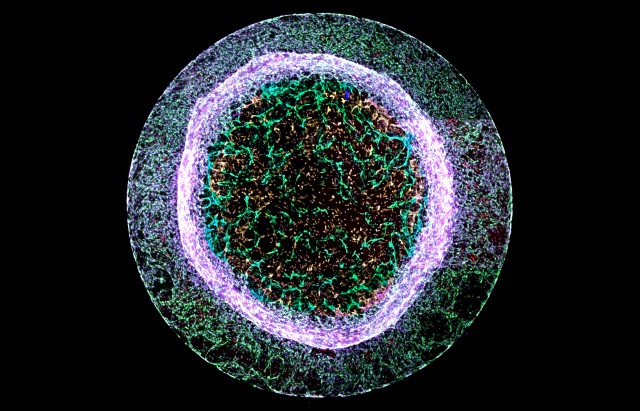
The colourful ‘Calcium rainbow’ won over the judges to receive the top prize. BHF-funded researcher, Dr Charlotte Buckley, took the image as part of her research into high blood pressure at the University of Strathclyde.
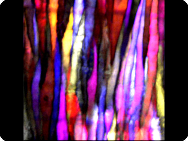
Resembling stained glass, this image shows the calcium signals of individual smooth muscle cells in an artery. Calcium is an important messenger that regulates the contraction and relaxation of blood vessels, which affects blood pressure and blood flow.
High blood pressure, also known as hypertension, is linked to various cardiovascular conditions, including stroke and vascular dementia. Although it is known that hypertension increases cardiovascular disease risk, the precise mechanisms behind this are poorly understood.
Using a 60x magnification water dipping lens at 10 frames per second for 1 minute, Charlotte is able to capture the time at which calcium signalling occurred, and colour-code them accordingly.
Blue indicates signals that took place at the start of the recording, running through purple, pink, red and orange, to yellow and white at the end of the recording.
By using this method, Charlotte hopes to better understand the role of calcium signalling in vascular health and disease and to find new ways to treat hypertension.
Dr Charlotte Buckley said: “I am thrilled the judges have chosen my image as the winner of the British Heart Foundations Reflections of Research image competition this year.
"This breathtaking image shows how art and science can merge in unexpected ways to highlight how research is not only beautiful, but also lifesaving.”
Dr Susanth Alapati’s entry, depicting an unexpected link between oral hygiene and heart health, captivated supporters to earn the title of Supporters’ Favourite.
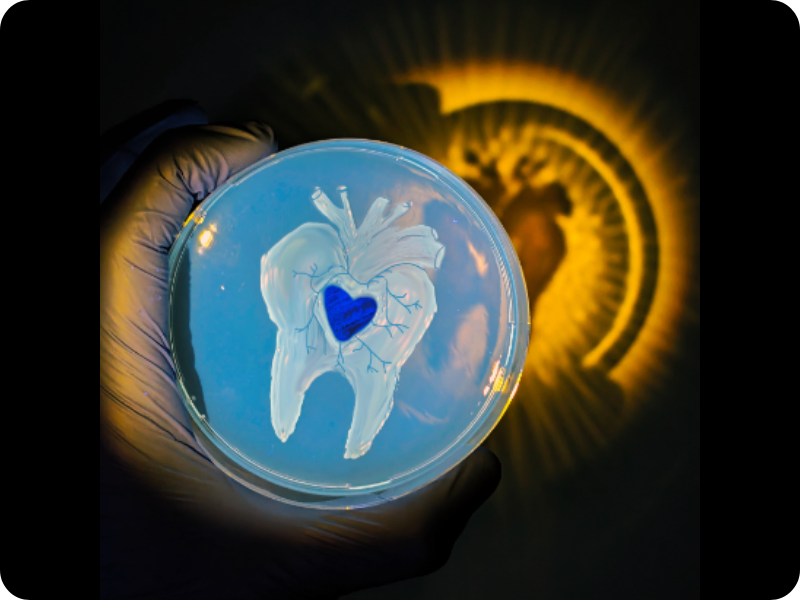
This artwork was created using bacteria in a Petri dish. The tooth structure was crafted using colonies of Staphylococcus bacteria – a typical inhabitant of the mouth – while the heart shape was formed from Porphyromonas colonies, a gum bacterium associated with gum disease.
This image emphasises the concept that inadequate oral hygiene, influenced by Staphylococcus and Porphyromonas, can potentially contribute to heart conditions like heart attacks and endocarditis – a dangerous infection of the heart lining and valves. These oral bacteria can enter the bloodstream when we chew, potentially causing inflammation in the arteries and heart.
Research consistently shows that oral health issues are connected to a higher risk of heart disease. Dr Alapati's research aims to better understand this connection in order to help doctors develop preventive strategies for patients.
Dr Susanth Alapati said: “I am delighted that this breathtaking image has been recognised by BHF supporters in the British Heart Foundation’s Reflections of Research competition.
"When it comes to keeping our hearts healthy, we often overlook the importance of healthy teeth and gums. Our research aims to uncover how certain gum bacteria in mouth adapt and contribute to heart attacks, helping us find better ways to prevent both gum and heart-related conditions.”

Paths of the heart’ wowed the judges and took home the top prize. Entered by BHF-funded researcher Dr Marina Strocchi, it was taken as part of her work at King’s College London that is now being continued at Imperial College London.
The multicoloured structure, generated by a computer model, is a map of bundles of cells that together make up the heart muscle. These bundles, each shown by a separate line, transmit the electrical signal that causes the heart to beat. The direction each line is pointing indicates the direction the electrical signal travels.
The signal starts in the two top chambers of the heart (the atria) and moves to the bottom two (the ventricles). By accurately modelling the heartbeat signal as it passes through the heart, Marina can simulate how an individual patient’s heart works, to better predict how they will respond to different treatments.
Dr Marina Strocchi said: “I am thrilled that my entry was chosen by the judges as this year’s winner. It is great to be able to share the beautiful side of my research.
“In our research, computer models help doctors understand so much more about diseases of the heart. By simulating how electrical activity spreads through the heart, following the direction shown in the picture, to then to trigger a heartbeat, we can predict how specific patients will respond to treatments to personalise and adjust how we care for them.”
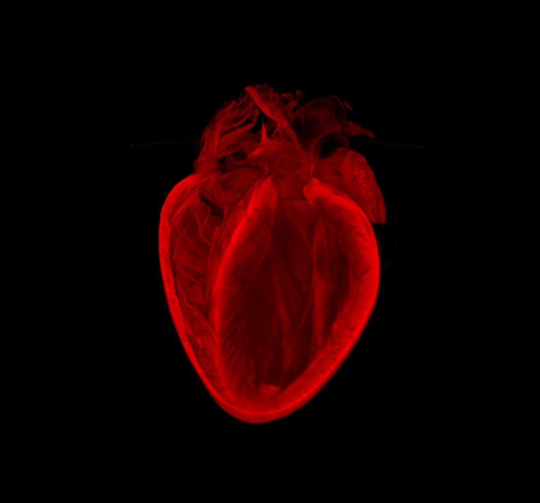
‘Seeing through the heart’, the image that captivated our supporters, was submitted by PhD student Judy Sayers. Fantastic bursts of light in this image highlight blood vessels around the outside of the heart and areas of the electrical system at its centre. Judy has made the heart transparent in the lab, exposing an explosion of activity in its electrical system that organises the heart’s rhythmic contraction to form a heartbeat.
It is this system that is visible as the tangle of fibres in the middle of the image. The two larger glowing areas are the atria – the top two chambers of the heart – and the branch around the outside on the left of the image, is a coronary artery, which supplies blood to the heart muscle.
Judy Sayers said: “I am delighted to have been recognised by the BHF’s supporters and it means a lot to be able to share with them the visual outputs of my research.
“Using chemical modification to make the heart transparent, we are able to view the heart’s dedicated electrical system in 3D without disturbing the anatomy by cutting the organ. This allows us to investigate how and why this system can fail in people with heart rhythm conditions. I hope that this research will lead to new therapeutic avenues for the treatment of these conditions.”
In 2022, ‘A flare of stellar vessels’, by Dr Régis Joulia, a BHF Research Fellow at the BHF Centre of Research Excellence, Imperial College London, took the win. Although we may think this is an image of a distant constellation or a tropical flower, it shows a small region of a human lung and its rich supply of blood vessels in red. The tiny green dots reveal immune cells called mast cells in the lungs and the blue dots are pericytes which are crucial cells for blood vessels to maintain their structure. Activation of the immune cells in chronic inflammatory conditions like asthma and chronic obstructive pulmonary disease can disrupt the structure of lung blood vessels, which can have detrimental effects on the amount of oxygen in the blood.
The winner of our 2020/21 competition was Dr Elisa Avolio, a post-doctoral research associate at the University of Bristol Medical School. Although at first glance it appears to resemble a luminous jelly fish, this image shows new blood vessel-like structures (pictured in green) sprouting from a 3D gel. These structures were created using a mixture of two types of heart cell - cardiac endothelial cells, which line the inside of every blood vessel, and pericytes, which ‘hug’ the outside of blood vessels to support the vessel and help it function. Encouraging new blood vessels to form after a heart attack to replace those that have died could help to re-establish blood supply to damaged areas of the heart and aid recovery.
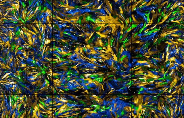
The 2019 winner 'A sea of cells' was created by Iona Cuthbertson at the University of Cambridge. What could be mistaken for the thick brushstrokes of a Vincent van Gogh painting bringing to life an ocean bloom, is in fact a close-up of smooth muscle cells that surround the blood vessels in mice. The smooth muscle cells, which are partly responsible for the control of blood flow by narrowing or widening blood vessels, are marked with differently coloured fluorescent proteins.
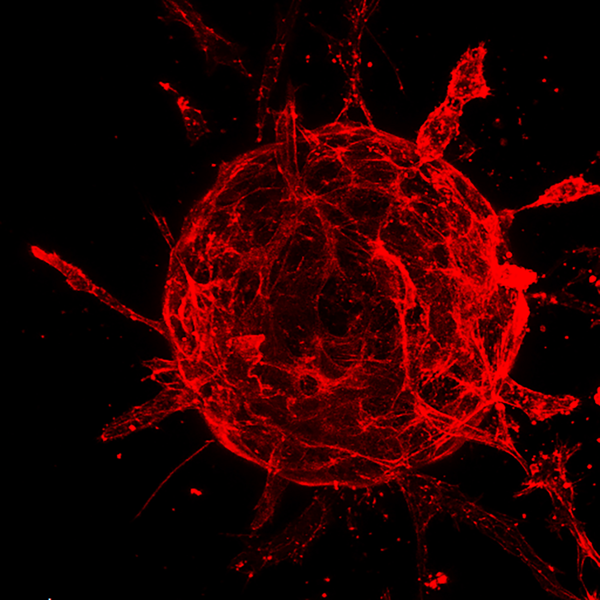
The 2018 winner ‘Explosive beginnings’ came from Courtney Williams, a Master’s and PhD student from the University of Leeds. The image shows a close-up snapshot of hundreds of endothelial cells – the cells which line all blood vessels – growing on the surface of a bead. They’re in the process of ‘sprouting’ which is the first step in the formation of new blood vessels.
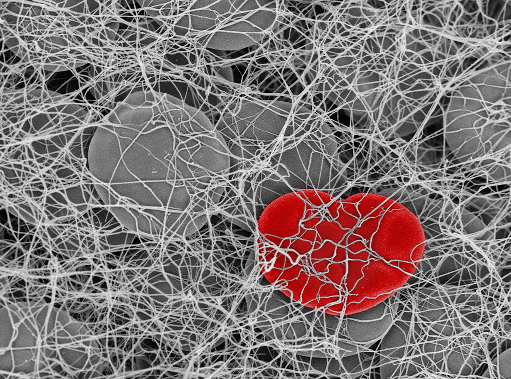
The winning image from 2017 came from Fraser Macrae at the University of Leeds titled ‘Getting to the heart of the matter’. The image takes us inside a deadly blood clot - the leading cause of heart attack and stroke. In the image red blood cells are trapped in the 3D mesh of fibrin fibres, which hold the clot together. One red blood cell had been compressed into a heart shape by the contracting fibres surrounding it.
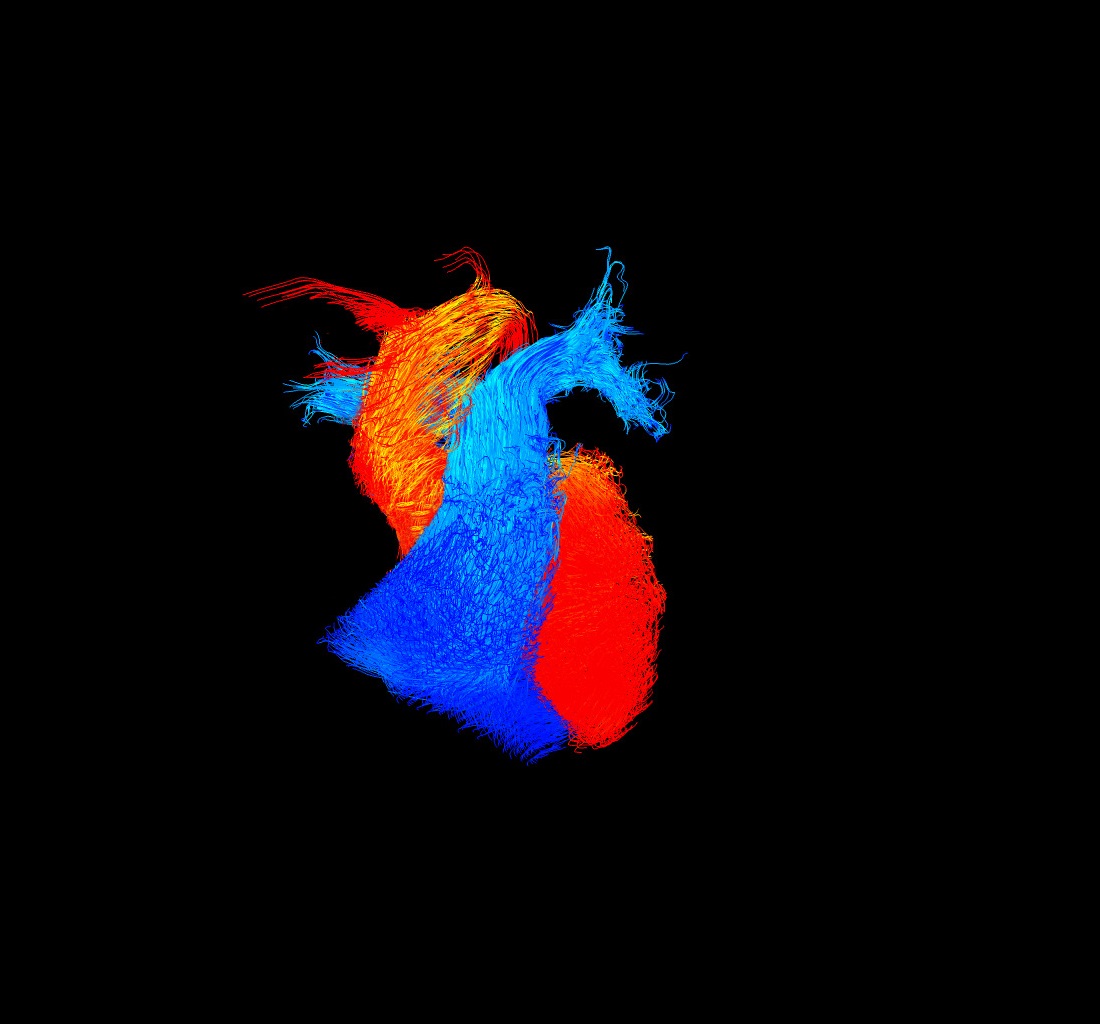
The winning image from 2015/16 came from Dr Victoria Stoll, from the University of Oxford. Titled 'Go with the flow', the image shows blood flow within the main pumping chambers of the heart - the ventricles - and through the main vessels leaving these chambers.
The blue flow is blood that needs oxygen, and is travelling towards the lungs, and the red is oxygen rich blood which has just passed through the lungs.
Victoria used this kind of image to visualise how patterns in blood flow are impacted by conditions like heart failure.
 This stunning winning image 'The clot thickens' came from Fraser Macrae at the University of Leeds. His picture of a blood clot has since been featured across the media and many of our publications and web pages.
The judges in 2014 were our Chief Executive Simon Gillespie; Roger Highfield, Director of External Affairs at the Science Museum, London; Daniel Glaser, Director of Science Gallery London; and Jasmine Pradissitto, a Quantum Artist.
Explore the whole shortlist in our Facebook album.
The judges in 2013 were James Gallagher, BBC Health and Science Reporter, our Medical Director Peter Weissberg and our Chief Executive Simon Gillespie.
The winning image came from a team at the University of Edinburgh, Gillian Gray, Megan Swim and Harris Morrison, with their image of the 3D structure of a mouse heart called The Broken Heart.
See the shortlisted images and videos from 2013 in our online album.
The winners of the 2012 competition accepted their prizes at the Annual Reception hosted at the Heartbreak Gallery.
The overall winner was a King’s College London team led by Dr Elisabeth Ehler, for her image B of the Bang, right. The Mending Broken Hearts award went to Ms Evie Maifoshie and colleagues at the Imperial College London.
See all of the shortlisted images and videos from 2012's competition in our Flickr album.

The winners of the 2011 competition accepted their prizes at a ceremony at the June conference of the British Cardiovascular Society in Manchester. As well as the usual image and video categories we launched a new prize in honour of our Mending Broken Hearts Appeal.
The overall winner was a Kings College London team led by Professor Nic Smith, for his image Feeding the Heart. left. The Mending Broken Hearts award went to Dr Renata Gomes and colleagues at the University of Oxford.
See all of the shortlisted images and videos from 2011's competition in our online album.
The winners of the 2009-10 competition were announced at a ceremony at the London Eye in February 2010 to coincide with National Heart Month. There were two categories: images and video.
The winner of the image category was Looking through the Heart by Dr Mathieu-Benoit Voisin and Miss Doris Proebstl from Queen Mary, right.
Dr Michael Markl, University of Freiburg, and Dr Philip Kilner, Imperial College, triumphed in the video category with their arresting movie of blood flows in the heart.
We made a film of the winners at the ceremony on London's South Bank.

We had an amazing selection of entries, and the overall winner was Dr Steve Thomas from the University of Birmingham for his stunning image of blood cells called 'platelets' being formed in bone marrow. left
He says the image "highlights their beauty and complexity". Dr Thomas is a postdoctoral research fellow in the laboratory of BHF Professor Steve Watson, a world expert in platelets.
Understanding more about platelets is vital to deciphering how blood clots are formed and broken down.

Richard Clayton from the University of Sheffield struck a chord with this image showing a computer simulation of electrical activity in the heart during the onset of cardiac arrest, right.
Instead of regular activity controlled by the pacemaker of the heart, electrical activation (shown in red) has looped around into a spiral that will fragment, resulting in lethal electrical anarchy.
“This type of computational model gives powerful insight into the mechanisms of normal and abnormal electrical activity in the heart while reducing the need for experiments using animal tissue.”

Aleksander Ivecic from Imperial College really impressed the judges with his story of cell migration.
"Cell migration could not be achieved without the presence of a cell skeleton (cytoskeleton). This reflects the way that the BHF has served to be the backbone of research investment in our department.
"The cell is migrating in a directed fashion which reflects a unified direction that every scientist under the BHF is making towards a better understanding of cardiovascular disease."
Find out more about our Reflections of Research imagery competition and how BHF-funded researchers can enter.
We have helped improve and save the lives of many people with heart and circulatory diseases. But these conditions still affect millions of families.
Over the past six decades, the BHF has supported important breakthroughs in our journey to beat heartbreak forever. Read the stories of our research successes.