Detailed map of the heart could lead to new treatments

Scientists we part-fund have created a cellular and molecular map of the healthy human heart, to understand how the organ functions, and to shed light on what goes wrong in heart disease.
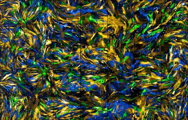
Researchers from all over the world analysed almost half a million individual cells to build a first cell atlas of the human heart. The atlas shows the huge diversity of cells and reveals heart muscle cell types, protective immune cells, and the intricate network of blood vessels. It also predicts how the cells communicate to keep the heart working.
Published in Nature, this study is part of the Human Cell Atlas initiative to map every cell type in the human body. The new molecular and cellular knowledge of the heart will enable better understanding of heart disease and guide more personalised medicine. It could also potentially lead to regenerative medicine in the future.
Shining a light on the heart's complexity
Our Associate Medical Director, Metin Avkiran, said: “Our hearts are fascinating and wonderfully complex organs made up of many different cell types. This ground-breaking study, which has been supported through a joint research funding scheme between the BHF and the German Centre for Cardiovascular Research (DZHK), has used cutting-edge technology to shine new light on that complexity in the healthy human heart.
"By mapping in exquisite detail the different cell types that reside in the human heart and the changes in their individual properties and interactions in disease, we can begin to identify better ways of preventing and treating many life-threatening conditions, from common rhythm disturbances such as atrial fibrillation to heart attacks and heart failure.”
Looking at half a million heart cells
The researchers discovered that there were major differences in the cells in different areas of the heart, and that each area of the heart had specific sets of cells, highlighting different developmental origins and potentially different responses to treatments.
Details never seen before
The researchers also studied the blood vessels running through the heart in unprecedented detail. The atlas showed how the cells in these veins and arteries are adapted to the different pressures and locations, and could help understand what goes wrong in the blood vessels during coronary heart disease.