Tests
What is an MRI scan?
Cardiac MRI has helped hundreds of thousands of people with heart problems over the last 40 years. Dr Phoebe Kitscha learns more from three British Heart Foundation-funded scientists who are making progress in this area.

Magnetic resonance imaging (MRI) is a type of medical scan first developed in the late 1980s. It uses a magnetic field and computer-generated radio waves to create detailed images of what’s going on inside the body.
Over the past 40 years, cardiovascular MRI (often called cardiac MRI, or CMR for short) has helped advance our understanding of heart and circulatory conditions, enabled the development of new treatments, and has become an important tool for diagnosis.

British Heart Foundation funding has played a vital role in getting us to this stage and has helped make the scanning process quicker and easier. Twenty years ago, it would take many minutes to capture a single still cardiac MRI image. Now, moving ‘cine’ MRI images of the heart can be captured in just a few seconds.
Today, per population, the UK has one of the highest number of CMR experts and hospitals offering it in the world. More than 100,000 CMR scans are done in NHS hospitals every year.
But what’s next for CMR? We share the perspectives of three BHF-funded researchers working in this rapidly developing field.
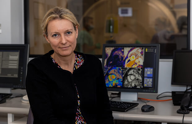
Charlotte Manisty (pictured above) is a Professor of Cardiology at University College London, and Consultant Cardiologist at Barts Heart Centre, where her focus is looking after people who develop heart problems as a result of cancer treatment.
While cancer survival in the UK has doubled in the last 40 years, some cancer drugs can have toxic effects on the heart.
Professor Manisty says: “The stakes are high for these patients. We don’t want to under-diagnose the effects the cancer treatment can have, as this can lead to irreversible heart damage. But over-diagnosing is risky too, as potentially lifesaving cancer treatment might have to be stopped.”
Today moving images of the heart can be captured in a few seconds
"We use CMR to monitor these patients as it’s the most precise measure we have of how the heart works, but importantly it also allows us to start learning more about how the damage is happening.”
She is leading a BHF-funded study looking at fluoropyrimidine chemotherapy, often used to treat gastrointestinal cancers. This treatment can cause heart problems such as chest pain, heart attacks and heart failure, but we don’t yet understand how this happens.
In this study, people starting fluoropyrimidine chemotherapy will have CMR scans before, during and after their treatment, looking for changes in the heart’s blood supply and inflammation in the heart muscle. It’s hoped this information can be used to develop ways to prevent and treat these effects.

In a second BHF-funded study Professor Manisty and her team are hoping to answer the question of what to do when someone who has developed heart problems as a result of cancer treatment then recovers after taking heart failure medication.
HER-2 therapy is used to treat certain types of breast cancer and can affect the heart’s pumping ability, but the heart usually recovers well after the therapy is finished. Most people in this situation don’t want to keep taking heart failure medication forever but at the moment, we simply don’t know whether it’s safe for them to stop.
In Professor Manisty’s study, 90 people whose heart has recovered after HER-2 therapy will be randomly assigned to either stop or continue their heart failure treatment. They will be carefully monitored with CMR scans.
Professor Manisty explains: “We’re using a ‘RapidCMR protocol’ that we’ve developed at Barts – a 13-minute scan which allows us to quickly assess how well the heart is pumping. The scans we previously did for cardio-oncology patients took 50 minutes, but this rapid scan means more people can benefit from the best possible monitoring.”
“Another area we’ve been working on is MRI in people with pacemakers or other implanted cardiac devices. Even though most of these devices are completely safe to go into a scanner, only about half of NHS Trusts will currently scan people who have them – either a heart scan or of any other part of the body. This is a real problem with equality in care, so we’ve done a lot of research around demonstrating that it’s safe and developing guidelines to help improve this.”
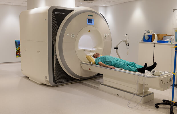
At the moment, a typical CMR scan takes 30 minutes to an hour. While these scans can give vital information to inform a person’s treatment, nobody enjoys being in a scanner for long periods of time. With the current pressures in the healthcare system and a shortage of cardiac radiographers, making the scanning process more efficient could help tackle growing waiting lists.
Making the scanning process more efficient could help tackle growing waiting lists
Dr James Howard, a BHF Intermediate Clinical Research Fellow and Consultant Cardiologist at Imperial College London, is hoping to use artificial intelligence (AI) to speed up the scan time for CMR. AI uses computers to mimic how the human brain identifies patterns and solves problems.
He is first focusing on a CMR technique called late gadolinium enhancement (LGE), used to look at areas of scarring in the heart – for example, as a result of a heart attack or problem with the heart muscle (cardiomyopathy). This technique is widely used but it can be a difficult type of scan to analyse and can be prone to ‘artefacts’ (where the scanner picks up a signal that isn’t really there). This means extra time has to be spent checking the images, and retaking them if needed, prolonging the scan time.
Dr Howard says: “My ambition is to get to a point where I could ask a medical student, who’s never seen an MRI scanner before, to click one button, and it will do the scan and tell us if the images are good enough for a diagnosis to be made.
“It’s not about replacing radiographers but making part of their job easier, and reducing the time people spend in the scanner. In some areas, people are waiting a year for a cardiac MRI. Streamlining the scan process with AI could help get more centres doing them, for more people.”
“MRI has really revolutionised our understanding of heart disease. Ten years ago, we used to talk about an inherited condition called arrhythmogenic right ventricular cardiomyopathy (ARVC). Now we call it arrhythmogenic cardiomyopathy (ACM), in part because with MRI we can see that often it doesn’t just affect the right side of the heart. I think the biggest change that will happen over the next ten years is speeding up scan times, and I would love my work to help us get to 15 minutes as the standard.”
Dr Oliver Rider is a BHF Senior Clinical Research Fellow and Associate Professor at the University of Oxford, where he leads a research group focused on using advanced MRI techniques to understand how the heart uses energy.
The pumping of blood by the heart is mostly powered by the heart muscle breaking down sugar and fatty molecules to produce an energy-carrying molecule called adenosine triphosphate (ATP). In a healthy adult heart ATP is mainly produced from breaking down fat, but it can switch to using more sugar for fuel when needed, such as during exercise. But during heart failure, the heart breaks down fewer fatty molecules and uses different energy sources less flexibly, producing less ATP, which reduces its ability to pump effectively.
These scans don’t just show us pictures but tell us more about how the heart is actually working
Dr Rider says: “The part of CMR that has always excited me is its ability to not just show us ‘pictures’ of the heart, but tell us more about how it’s actually working. We use CMR and a technique called magnetic resonance spectroscopy (MRS) to learn more about the balance between fats and sugars for energy production in the heart, and how this relates to how the heart is working.”
In his project, Dr Rider is using MRS to help understand how changing the source of energy the heart is using could help tackle heart failure. One part of the study is looking at a drug called fenofibrate, which is sometimes used as a cholesterol-lowering treatment and helps boost the breakdown of fat molecules. People with heart failure will be randomly assigned to receive fenofibrate or not, to see how this affects the heart’s energy use, and whether this cheap drug could be repurposed to help improve heart failure symptoms.

The second part of the study is focused on learning how the heart’s use of energy changes in people with heart failure with preserved ejection fraction. This is a relatively under-investigated form of heart failure, where the heart contracts as normal but can’t relax and fill with blood properly between beats.
He says: “Compared to other ways of imaging the heart, you get so much more information from CMR. In one stop, you can look at all the heart’s structures. You can see how the heart works, its blood supply, valves, inflammation and scarring. That gives you clearer and more useful answers to medical questions.
“The difference between the scans now and when I started at Oxford in 2005 is a real step up in terms of image quality and what information we can get. BHF support for CMR research has really helped with driving advances.”
Supported by BHF funding, his team has helped develop a technique called hyperpolarised MRI, which boosts MRI signals 10,000-fold. He says: “This gives us the ability to pick up changes in the heart’s energy use that were simply impossible to detect before. This will help us understand heart disease better and pave the way for new treatments.”